• Home
    • Astronomy Photos
    • Chemistry Photos
    • Climate Change Photos
    • Planet Earth
    • Earth Science Images
    • Earthquakes
    • Galaxies & Nebulae
    • Geology
    • Infinity Images
    • Meteorology Images
    • Microbiology Images
    • Micrographs
    • Nanotechnology
    • Night Sky
    • Noether Symmetry
    • Physics Images
    • Research & Technology
    • Space Illustration
    • Tornado Images
    • X-ray Art
    • Medical Micrographs
    • Medical Photos
    • Psychology Images
    • Famous Actors
    • Famous Artists
    • Famous Music Composers
    • Famous Musicians
    • Famous Scientists
  • Medical Video
    • Company Info
    • Contact Us
    • FAQ
    • Custom Design
  • Pharma Images
  • Science & Medical Blog
Menu

Science & Medical Stock Images | Science Source

Stock Medical Images, Medical Illustration | Science Source
  • Home
  • Science Photos
    • Astronomy Photos
    • Chemistry Photos
    • Climate Change Photos
    • Planet Earth
    • Earth Science Images
    • Earthquakes
    • Galaxies & Nebulae
    • Geology
    • Infinity Images
    • Meteorology Images
    • Microbiology Images
    • Micrographs
    • Nanotechnology
    • Night Sky
    • Noether Symmetry
    • Physics Images
    • Research & Technology
    • Space Illustration
    • Tornado Images
    • X-ray Art
  • Medical Images
    • Medical Micrographs
    • Medical Photos
    • Psychology Images
  • Culture Images
    • Famous Actors
    • Famous Artists
    • Famous Music Composers
    • Famous Musicians
    • Famous Scientists
  • Medical Video
  • About Us
    • Company Info
    • Contact Us
    • FAQ
    • Custom Design
  • Pharma Images
  • Science & Medical Blog

The Power of Tornadoes: Exploring Supercells, Tornado Formation, and Climate Change

April 25, 2024

Tornadoes emerge from the tumultuous core of thunderstorms, leaving destruction in their wake. Understanding tornadoes necessitates diving into the complexities of supercells, where these vortexes are born. We will delve into the science behind tornado formation while also considering the influence of climate change on these extreme weather events.

Gallery of Tornadoes and Supercells

Supercell Anatomy

Supercell thunderstorm, Kansas. © Caryn Hill Photography / Science Source

Supercells stand out from thunderstorms for their rotating updrafts or mesocyclones. These colossal atmospheric systems, which span vast distances and persist for hours, are the primary incubators for tornadoes. A supercell starts with the clash of warm, moisture-laden air rising from the Earth's surface with cooler, drier air aloft. This stark thermal and moisture gradient creates an unstable atmosphere conducive to thunderstorm development.

As the warm, moist air ascends, it undergoes rapid condensation, releasing latent heat and fueling the updraft's ascent. Concurrently, horizontal wind shear—variations in wind speed and direction with altitude—lays the groundwork for rotational motion within the storm. The interaction between the updraft and wind shear initiates the formation of the mesocyclone, a rotating column of air several miles in diameter.

A Tornado Begins to Form

While supercells possess the necessary ingredients for tornado formation, not all lead to the creation of tornadoes. The transition from a rotating mesocyclone to a tornado involves the interplay between the storm's internal dynamics and external environmental factors. One pivotal mechanism driving tornado genesis is the stretching of horizontal vorticity—a spinning motion of air—into a vertical orientation.

This vertical stretching occurs through various processes, including the tilting of horizontally rotating air by the updraft and the convergence of horizontal wind vorticity near the Earth's surface. As the mesocyclone intensifies, a wall cloud—a region of enhanced low-level rotation—may form beneath the storm. A funnel cloud may descend from this wall cloud, marking the incipient stage of a tornado. The funnel cloud extends downward from the parent cloud base but does not yet contact the ground.

Tornado Formation

Tornado in four steps. © Monica Schroeder / Science Source

A critical process known as vortex stretching must ensue for a funnel cloud to evolve into a tornado. Vortex stretching amplifies the rotation within the funnel cloud, intensifying its circulation and drawing air inward at an accelerating rate. As the rotating column of air descends toward the Earth's surface, it may become visible as a tornado. Upon contact with the ground, the tornado establishes itself, unleashing its destructive potential.

Waterspouts

Historical Drawing of a Waterspout. © Mary Evans / Science Source

Waterspouts are the aquatic counterparts of tornadoes. They form over bodies of water, ranging from oceans and lakes to rivers and even smaller ponds. While waterspouts share similarities with tornadoes, their formation mechanisms differ slightly. Waterspouts typically develop under high humidity and atmospheric instability conditions, often associated with convective showers or thunderstorms passing over warm water. Like their terrestrial counterparts, waterspouts exhibit a characteristic funnel shape, extending downward from the cloud base toward the water's surface. While most waterspouts remain relatively weak and short-lived, some can intensify, posing risks to maritime activities and coastal communities. 

The Fujita Tornado Scale

With winds of between 250 and 330 km/h, is an F3 tornado © QA International / Science Source

The Fujita Tornado Scale, developed by renowned meteorologist Dr. Tetsuya Theodore Fujita in 1971, is a vital tool for categorizing tornadoes based on their intensity and damage. Originally known as the Fujita-Pearson Scale, it underwent revisions over the years, with the Enhanced Fujita (EF) Scale being the most recent iteration adopted in 2007. Based on estimated wind speeds and associated damage patterns, this scale classifies tornadoes into six categories, ranging from EF0 (weak) to EF5 (violent). Each category provides insights into the destructive potential of a tornado, helping meteorologists and emergency management officials assess the severity of a tornado event and allocate resources accordingly. The EF Scale facilitates standardized reporting and analysis of tornado events and enhances public awareness and preparedness, empowering communities to respond effectively to the ever-present threat of tornadoes.

Effects of Climate Change

 In recent years, mounting evidence shows climate change influences the frequency, intensity, and distribution of tornadoes. Increasing atmospheric moisture and changes to circulation patterns due to climate change shape tornado occurrence.

Gallery of Weather and Meteorology Images

Our Future in the Face of Tornadoes

Computer forecasting extreme weather. © Andrew Baker / Science Source

Tornadoes are formidable manifestations of atmospheric dynamics, embodying nature's raw power and complexity. While scientists continue to advance our understanding of tornado formation, the potential influence of climate change underscores the urgency of addressing environmental challenges. By combining scientific inquiry with proactive mitigation and adaptation strategies, we can strive to mitigate the impacts of tornadoes and build resilient communities in the face of evolving climatic conditions. As we marvel at the spectacle of these swirling vortexes, let us also heed the call for collective action to safeguard lives and livelihoods from the ever-changing forces of nature.

Tags tornadoes, weahter, supercell, stock images, science
Comment

Microscope photo of head lice. © STEVE GSCHMEISSNER / Science Source

The Microscopic Metropolis Inhabiting Your Skin: Mites, Lice, Fleas, Bacteria and Fungus

April 18, 2024

As we gaze into the mirror, we often overlook the bustling metropolis that thrives beneath the surface – our skin. Beyond its protective and aesthetic functions lies an ecosystem teeming with microscopic inhabitants. Join me on an expedition into this unseen realm as we unravel the mysteries of the creatures that call our skin home.

Follicle mite protruding from a hair follicle. © Andrew Syred / Science Source

Demodex folliculorum and Demodex brevis: Eyelash Mites

Meet the microscopic mites that have taken up residence within the confines of our hair follicles – Demodex folliculorum and Demodex brevis. These microscopic arachnids, measuring a mere fraction of a millimeter, spend their entire lives deep within our skin's sebaceous glands and hair follicles. While Demodex folliculorum primarily inhabits the hair follicles, Demodex brevis prefers the sebaceous glands. Despite their benign presence for most individuals, these mites have been implicated in certain skin conditions when their populations increase unchecked, leading to symptoms such as rosacea and blepharitis.

Human Lice: Head Lice, Body Lice & Public Lice

The mention of lice conjures images of scalp discomfort, but these parasites can also be found in other areas. Three distinct species of lice – head lice (Pediculus humanus capitis), body lice (Pediculus humanus corporis), and pubic lice (Pthirus pubis) – have adapted to specific niches on the human body. Head lice primarily inhabit the scalp and hair. Body lice prefer clothing and bedding, venturing onto the skin only to feed. Pubic lice prefer coarse body hair, particularly in the pubic region. These parasites evoke both discomfort and social stigma.

Human flea (pulex irritans). © Eye of Science / Science Source

Human Fleas

Fleas, notorious for tormenting household pets, are not averse to seeking refuge in human hosts. Human fleas (Pulex irritans) can infest areas lacking hygiene, increasing in densely populated and unsanitary environments. While they primarily feed on the blood of mammals, including humans, their presence can provoke allergic reactions and transmit diseases such as murine typhus and tapeworm infections. Vigilance in maintaining cleanliness and addressing infestations promptly is essential for mitigating the risks associated with these persistent pests.

Staphylococcus epidermidis bacteria, the cause fo a staph infection. © Steve Gschmeissner / Science Source

Skin Bacteria

Our skin is a veritable battleground for microbial communities, with bacteria comprising many of its inhabitants. While many bacterial species contribute to maintaining skin health and immune function, certain strains can instigate infections and dermatological disorders. From the ubiquitous Staphylococcus aureus to the opportunistic Propionibacterium acnes, the intricate interplay between bacterial populations shapes the resilience and susceptibility of our skin to disease.

Gallery of Acne Medical Photographs and Medical Illustrations

Skin Fungus: Ringworm, Athlete's Foot

Illustration of fungus Trichophyton rubrum and athlete’s foot. © Kateryna Kon / Science Source

Fungal infections are another common affliction of the skin. Common ones are ringworm and athlete's foot. Dermatophytes, the fungi responsible for these conditions, thrive in warm, moist environments, making areas such as the feet, groin, and scalp prime targets. Despite their colloquial names, neither ringworm nor athlete's foot is caused by worms; instead, they are fungal.

Our skin is a dynamic ecosystem inhabited by microscopic organisms, each playing a distinct role in its equilibrium. From the elusive Demodex mites to the persistent parasites and opportunistic pathogens, understanding the complexities of our skin's inhabitants enhances our appreciation for its resilience and vulnerability. Fostering a harmonious relationship becomes essential in preserving the health and integrity of our largest organ.

Tags health, medical, eyelash mites, lice, stock images
Comment

Scanning electron micrograph (SEM) of a water bear (Macrobiotus sapiens) in moss © Eye of Science/Science Source

Exploring Microscopy: Ultimate List of Microscope Types With Image Examples

April 10, 2024

A glance at any scientific or medical article reveals how far microscopy has evolved from the basic microscope of high school biology classes. Today, it's not just about magnification but also about capturing sharp details, enhancing depth of field, observing internal structures, and experiencing vivid 3D-like visuals that captivate our imagination. Over time, there have been numerous advancements in light microscopy, introducing many new types that unveil a previously unimaginable level of detail.

Stock Image Gallery of Medical Micrographs

The conventional microscope, which is familiar from childhood and known as the "light microscope," functions by utilizing light and a series of lenses. By incorporating filters, specialized mirrors, lasers, specific light spectrums, and other enhancements, we can extract significantly more detailed information.

First, let's cover a few basics:

What is Microscopy

Electron micrograph of red blood cells and fibrin. © Scoatt Camazine / Science Source

Microscopy could be defined as the scientific technique used to observe objects that are too small to be seen with the naked eye, typically using microscopes. It allows researchers to examine the structure and properties of materials and biological specimens at a microscopic level.

Magnification and Resolving Power

Magnification refers to the degree to which an object is enlarged when viewed through a microscope. It is usually expressed as a ratio (e.g., 100x), indicating how many times larger the object appears compared to its actual size.

Resolving power (or resolution) refers to the microscope's ability to distinguish between two closely spaced objects as separate entities. It determines how clearly individual details can be seen.

A Gallery of Royalty-free Micrographs

Let's start with Light Microscopes:

Four types of Light micrographs: Bright Field, Dark Field, Polarized, Phase Contrast. © Marek Mis/Science Source

Various illumination techniques are employed when examining slides under a light microscope, including Bright Field, Dark Field, Fluorescence, Differential Interference Contrast (DIC), Phase Contrast, and Confocal microscopy.

Photos of the microscopes can be found below.

Light Microscope Using Bright Field

Bright Field Microscopy: The light source is directed beneath the specimen, resulting in a brightly lit area surrounding it.

Light Microscope Using Dark Field

Dark Field Microscopy: Here, the light source is obscured, casting light onto the specimen from various angles, providing slightly different details compared to direct illumination.

Fluorescence Microscope

Fluorescence Microscopy: This technique utilizes light filters and specific wavelengths to induce fluorescence in the specimen, producing vibrant images.

Purkinje neurons of the cerebellar cortex stained with four staining methods: hematoxylin eosin (top left), cresyl violet (top right), Cajal's silver nitrate (bottom left) and silver method for Golgi apparatus (bottom right). © Jose Luis Calvo/Science Source

Phase Contrast Microscope

Phase Contrast Microscopy: By employing specialized lenses and filters, this method allows the visualization of transparent and colorless specimens with minimal shadowing.

Differential Interference Contrast Microscope

Differential Interference Contrast Microscopy (DIC): Through the use of polarizers, beam splitters, condensers, and filters, transparent specimens can be observed with a distinctly three-dimensional appearance.

Different illumination techniques of a light microscope: dark field, fluorescence, bright field, phase contrast, DIC (differential interference contrast). Child's hair strand © Ted Kinsman/Science Source

Caenorhabditis elegans (roundworm) embryo transform into a Dauer state when stressed during larval development (for instance, due to lack of food, hot environment, or crowding). Somatic cells are colored red, and yellow dots show gut granules used for fat storage. Image compiled from scans of different layers of the embryo. © Kul Bhatia / Ming Kin Wong / Science Source

Confocal Microscope

Confocal Microscopy: Utilizing Confocal Laser Scanning Microscopy (CLSM), a laser and spatial pinhole combine to produce high-resolution images with enhanced clarity.

Gallery of Botany Micrographs

Advanced types of microscopes:

Scanning electron micrograph (SEM) of Escherichia coli (E. coli), a type of Gram-negative rod-shaped bacteria that normally lives in the intestines of people and animals. Magnification is 17,500x at 15cm image size. ©Nano Creative / Science Source

Scanning Electron Microscope

Scanning Electron Microscopy (SEM): This microscope utilizes a beam of electrons to generate sharply detailed images by detecting reflected electrons off the specimen's surface within a vacuum environment, offering magnifications ranging from 20x to 30,000x with a spatial resolution of 50 - 100nm.


Transmission electron micrograph (TEM) of bacteriophage. © Biophoto Associates / Science Source

Transmission Electron Microscope

Transmission Electron Microscopy (TEM): By passing a beam of electrons through a thinly sliced specimen, TEM reveals intricate internal structures with magnifications of up to 2,000,000x.


Plasma membrane proteins. Colored atomic force micrograph (AFM) showing the surface of a plasma membrane. The membrane consists of a lipid bilayer with many proteins (peaks) attached to it. © PROF. DR. H.OBERLEITHNER, UNIVERSITY HOSPITAL OF MUENSTER / Science Source

Atomic Force Microscope

Atomic Force Microscopy (AFM): AFM operates by bouncing a laser off a stylus attached to a cantilever lever, which then traces the specimen's surface. Any deviations trigger sensors, generating a raster image that includes the Z-Plane. Unlike electron microscopy, AFM doesn't require a vacuum, offering versatility in various environments.


False-color scanning tunneling micrograph (STM) of a DNA double-helix; approximately three turns of the helix are displayed in this image. © Driscoll, Youngquist, & Baldeschwieler / Caltech / Science Source

Scanning Tunneling Microscope

Scanning Tunneling Microscopy (STM): STM employs electrons based on quantum tunneling, allowing for atomic-level surface imaging in vacuum, air, water, or ambient gas environments.


What can we look at with all of these microscopes?

Scanning Electron Micrograph (SEM) of unidentified freshwater bacteria collected from pond water. © Ted Kinsman / Science Source

Scoop up pond water or ocean water to be astonished by the plethora of living zooplankton and phytoplankton visible within a single drop using a simple light microscope.

Gallery of Phytoplankton and Zooplankton

It opens you to the wonder of cyanobacteria, blue-green algae, and ciliates like paramecium, daphnia, amoebas, and euglena. If you were lucky, you might have witnessed them conjugate and divide!


Diatom (Stephanopyxis sp.) from a South Atlantic Ocean deep sea probe. Scanning electron microscope. © Eye of Science / Science Source

Additionally, ocean water drops allow a peek at copepods, immature mollusks, krill, algae, crustaceans, and fish in their zooplankton stage. You may also behold the breathtaking beauty of diatoms, the most common type of phytoplankton in our oceans.

Switch to a higher powered Scanning Electron Microscope (SEM) to view Water Bears, pollen, blood cells, and insects. An SEM uses a particle beam of electrons to photograph the surface of a vacuum-sealed specimen.

Transmission Electron Microscopes (TEM) allow us to see cross-sections of specimens, such as the beautiful interior of the human body, marine life, and animal and plant cells. The TEM's particle beam passes through its vacuum-sealed specimen.

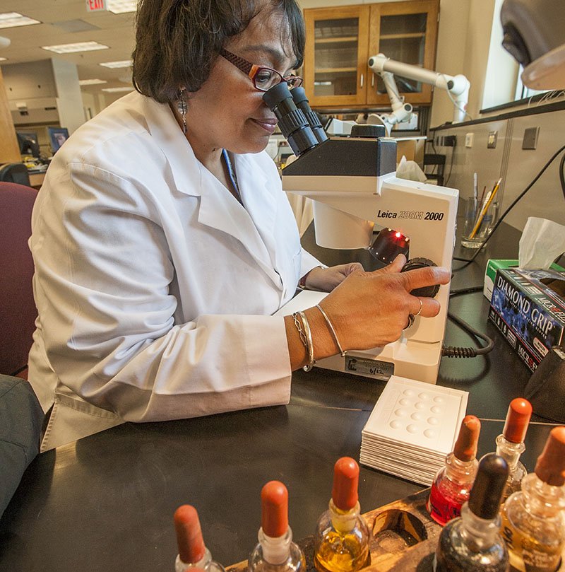
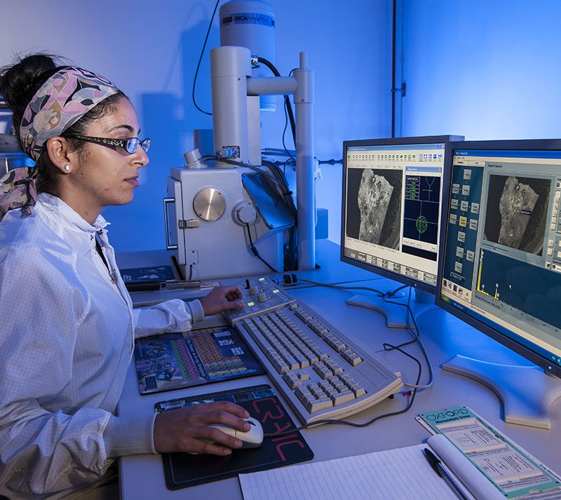
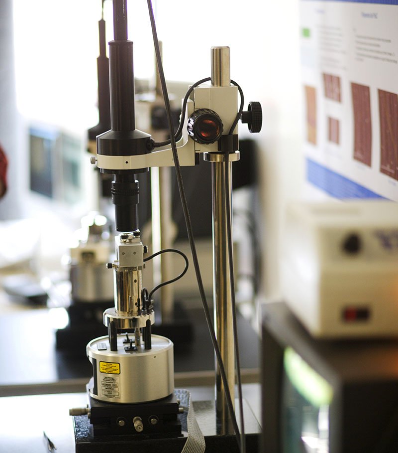
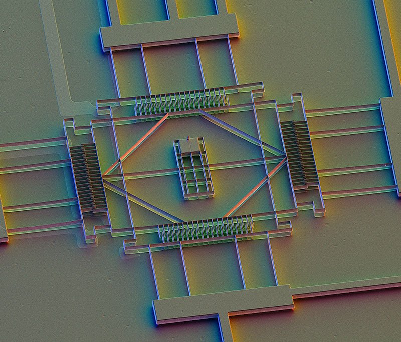
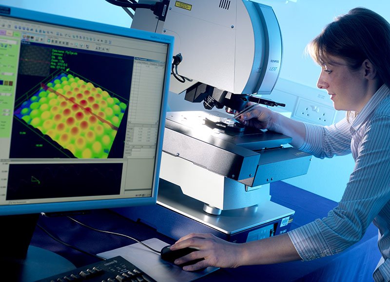
Photos of Types of Microscopes More Photos of Microscopes

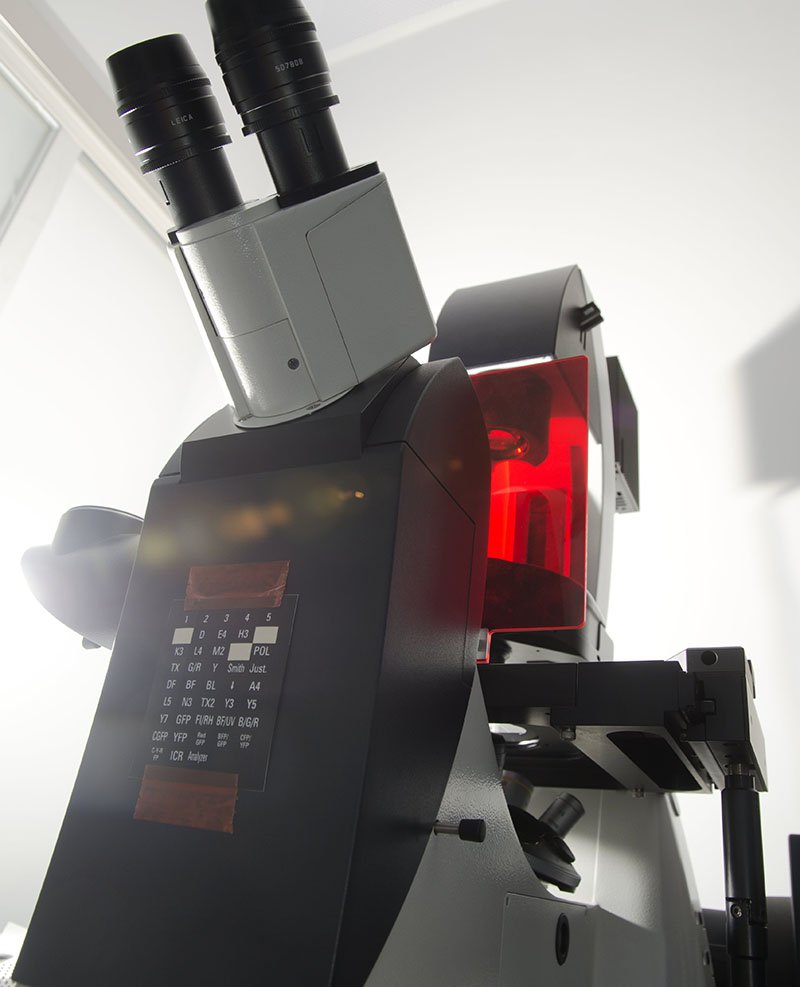
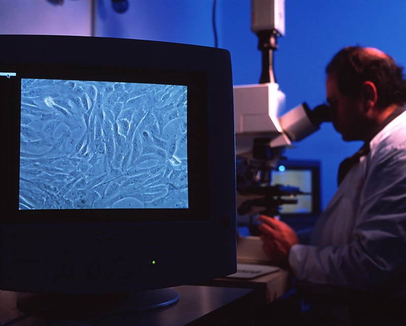
Light Microscope
Light Microscope
Scanning Electron Microscope
Atomic Force Microscope
Scanning Tunneling Microscope STM
Florescence Microscope
Phase Contrast Microscope
Confocal Scanning Microscope
Transmission Electron Microscope
Gallery of Microscope Types

Applications of medical microscopy

Bone marrow cell. This micrograph shows this cell with light (inset) and electron microscope. © Jose Luis Calvo/Science Source

We can view the cells of the human body with many different microscopes. The light mic, SEM, and TEM show scientists and medical researchers different angles and aspects of the cell and its fine structures and organelles.

Microscopes help scientists study cancer - breast, ovarian, prostate, liver, and skin cancer. We can better understand skin conditions such as psoriasis and eczema. They assist in the fight against nervous, respiratory, and circulatory system diseases. And a cure for muscular conditions such as fibromyalgia and multiple sclerosis (MS), and autoimmune conditions.

The Future of Microscopy:

Microscopy methods continue to evolve, with innovations like Acoustic Microscopy, which utilizes sound waves akin to SONAR, promising exciting prospects for the future of scientific exploration.


Tags microscopes, microscopy, scanning, electron, stock images, stock photos
Comment

Visual History of Medicine: Ancient Civilizations Through the 20th Century

January 23, 2024

The history of medicine is woven with ancient wisdom, scientific breakthroughs, and human curiosity. From the humble beginnings of ancient herbal remedies to the modern invention of pharmaceuticals, the evolution of medicine mirrors the progress of humanity itself. Let’s explore some of the pivotal moments and developments that have shaped healthcare.

VIEW THE MEDICAL HISTORY GALLERY OF STOCK IMAGES

Ancient Herbal Remedies:

A photos of the Ebers medical papyrus from ancient Egypt. © Science Source

The roots of medicine stretch back to the dawn of civilization, where early humans relied on nature for healing. Ancient herbal remedies, passed down through generations, formed the cornerstone of early medical practices. Civilizations such as the Sumerians, Egyptians, and Chinese meticulously documented the therapeutic properties of plants, laying the foundation for botanical medicine.

Egyptian Medicine:

In the land of the Pharaohs, medicine flourished alongside advancements in architecture, astronomy, and art. Ancient Egyptian physicians, revered for their expertise, blended empirical observation with spiritual beliefs to diagnose and treat illnesses. The Ebers Papyrus, a monumental medical document dating back to around 1550 BCE, provides insights into the ailments and remedies of the time, ranging from surgical procedures to herbal concoctions.

Ancient Greek Medicine:

The legacy of Hippocrates, the father of Western medicine, looms large over the annals of ancient Greek medicine. Rejecting superstition in favor of rational inquiry, Hippocratic physicians laid the groundwork for clinical observation and ethical medical practice. The Hippocratic Oath, a timeless pledge to uphold patient welfare and confidentiality, remains a guiding principle for modern healthcare professionals.

Medieval Medicine:

A lamb on a table is strapped to the arm of a man so that the lamb's blood is transfused into him.

Early blood transfusion. Historical artwork of the English physiologist Richard Lower (1631-1691) transfusing blood into a man's arm from a lamb in 1667. The end of the tube used to puncture the blood vessels and transfer the blood is at top left. ©Science Source

The Middle Ages ushered in an era of profound religious influence and medical stagnation. Drawing from the teachings of Galen, medieval physicians adhered to the theory of the four humors, believing that an imbalance in bodily fluids caused disease. Monastic infirmaries and Islamic medical centers served as oases of healing amidst the turmoil of war and plague, preserving and transmitting ancient medical knowledge to future generations.

Renaissance Medicine:

The rebirth of learning during the Renaissance sparked a resurgence of interest in anatomy, physiology, and empirical investigation. Visionaries such as Leonardo da Vinci and Andreas Vesalius revolutionized the study of human anatomy through meticulous dissections and anatomical illustrations. The printing press, a transformative invention of the era, disseminated medical texts to a wider audience, democratizing access to knowledge.

19th Century:

The 19th century witnessed unprecedented strides in medical science and public health. The discovery of anesthesia by William Morton in 1846 revolutionized surgical practice, paving the way for complex procedures with reduced pain and risk. The germ theory of disease, championed by Louis Pasteur and Robert Koch, fundamentally altered our understanding of infectious diseases, leading to advancements in sanitation, vaccination, and antibiotics.

20th Century:

The dawn of the 20th century heralded a golden age of medical innovation and global health initiatives. The development of insulin by Frederick Banting and Charles Best transformed the prognosis for diabetes patients, while the discovery of penicillin by Alexander Fleming inaugurated the era of antibiotics. Breakthroughs in medical imaging, genetics, and pharmacology reshaped diagnostic and therapeutic modalities, offering new hope for patients facing previously incurable conditions.

Modern Drugs:

The advent of modern pharmaceuticals represents a triumph of science and collaboration. From aspirin to statins, from antiretrovirals to immunotherapies, pharmaceuticals have revolutionized disease management and prevention, extending lifespans and enhancing quality of life for millions worldwide. However, the pursuit of medical progress is not without challenges, as concerns over drug safety, affordability, and equitable access persist in an increasingly interconnected world.

The Journey of Medical History

The history of medicine is a testament to human ingenuity, resilience, and compassion. From ancient herbal remedies to cutting-edge pharmaceuticals, each chapter in this narrative reflects our relentless quest to alleviate suffering and prolong life. As we stand on the threshold of a new era in healthcare, let us honor the lessons of the past while embracing the opportunities of the future, united in our commitment to heal, to innovate, and to care.

Tags history, medicine, medical, stock images
Comment

Featured Posts

Featured
Dec 2, 2024
CRISPR Gene Editing Technology: Bringing Hope for the Future
Dec 2, 2024
Dec 2, 2024
Oct 7, 2024
New Hope for Sickle Cell Disease: Innovative Treatments on the Horizon
Oct 7, 2024
Oct 7, 2024
May 12, 2024
Historical Portrait Collection: A Resource for Education
May 12, 2024
May 12, 2024
Apr 25, 2024
The Power of Tornadoes: Exploring Supercells, Tornado Formation, and Climate Change
Apr 25, 2024
Apr 25, 2024
Apr 18, 2024
The Microscopic Metropolis Inhabiting Your Skin: Mites, Lice, Fleas, Bacteria and Fungus
Apr 18, 2024
Apr 18, 2024
Apr 10, 2024
Exploring Microscopy: Ultimate List of Microscope Types With Image Examples
Apr 10, 2024
Apr 10, 2024
Feb 20, 2024
The Fifth Solvay Conference: an Exclusive hand-colored Stock Photograph
Feb 20, 2024
Feb 20, 2024

CUSTOM ILLUSTRATION DESIGN         MEDICAL STOCK IMAGES          SCIENCE STOCK IMAGES

STOCK HISTORY IMAGES          ENVIRONMENTAL IMAGES      BUY PRINTS

All Contents © 2025 Science Source Stock Images 307 Fifth Avenue, New York, NY 10016  212-758-3420